English

Treatment

Integrated treatment of cleft lip and palate

There are various problems due to cleft lip and palate during the growth process.
Appropriate treatment at the appropriate timing can reduce the negative influence to the growth affected by cleft lip and palate. This hospital is a specialized dentistry hospital, which is rare in Japan. Therefore, there are many specialists in various fields, and there is also a diversity that can not been seen in other hospitals. Experts in each department can provide the best treatment from their respective positions.

Also, daily research is being conducted with the aim of further improving treatments.
Depending on age, there might be negative feelings to having a surgery. However, children who are raised with love by those around them will be able to go out into the world with confidence both physically and mentally when they grow up. We strive each day to provide assistance.

  • Cleft lip
  • Cleft lip and palate
  • Cleft palate
Birth
First visit
Breastfeeding instruction
Impression talking *
Set a Hotz plate *
3~6 months old
Cheiloplasty
1 year old
Palatoplasty (closure of soft palate)
Start of speech management *
Caries treatment and oral management *
1 year and 6 months old
Palatoplasty (closure of anterior of hard palate)
2 years old
3 years old
Start of speech treatment *
4 years old
Start orthodontic treatment
Over 5 years old
Bone graft
6 years old
Minor rhinoplasty or lip repair
Over 15 years old
Rhinoplasty and secondary lip repair
Over 18 years old
Orthognathic surgery
Around 19 years old
Pharyngeal flap surgery
Prosthetic treatment
End of treatment

* Treat as needed

Problems due to cleft lip and palate

There are many problems caused by cleft lip and palate. Thus, it is necessary to perform the appropriate treatment at the appropriate time to overcome these problems. These problems are as follows:

  • 1
    Decreased feeding ability
  • 2
    Aesthetic problems
  • 3
    Speech impairment
    (impairment of pronunciation of words)
  • 4
    Developmental disorders of the maxilla
  • 5
    Malalignment of teeth
  • 6
    Exudative otitis media

1 Decreased feeding ability

In order for the baby to catch the nipple and drink the milk, it is necessary for the lips, upper jaw, lower jaw, tongue, etc., to work cooperatively to repeatedly perform the action of pressing the nipple and the action of forming negative pressure in the mouth and sucking (sucking the nipple) alternately.

For children with cleft lip and palate, it is difficult to press the nipple because of the cleft in the upper jaw, and even if one attempts to make the pressure in the mouth negative, air will come in from the nose and the ability to suck the nipple becomes extremely weak. Thus, it is necessary to provide much assistance. We recommend using a Hotz-type plate and nipple designated for cleft palate. (ChuChu cleft palate nipple, Pigeon P nipple, etc.). A Hotz-type plate is a device like a denture that covers the cleft part of the upper jaw. It is adjusted and remade as the patient grows, and it can be used until about the age of one year. This plate helps to improve feeding and also regulates maxillary growth to create a better maxillary shape.

Recently, an NAM-type plate has been developed to cure deformities of the nose and lips, even a little before surgery, and has been adopted at this center.

It may take a few weeks before the baby can drink well, so remain confident and patient. Since it is easy for the baby to swallow air while using this plate, hold the baby in a slightly upright position and perform feeding while checking the tension of the stomach from time to time. When the stomach feels tense, take a break and burp the baby. As a guide, the feeding time should be within 20 minutes. If possible, massage the breasts and breastfeed the baby. After feeding, remove the plate from the mouth and wash it with running water. Breastfeeding directly without the plate is difficult to get enough nutrition because it is difficult to suck out milk, but it is acceptable to perform it for the sake of mother-child contact. It is necessary to visit the hospital once every 1-2 weeks until the age of about one year to adjust the plate.

Hotz-type plate

Hotz-type plate

NAM-type plate

NAM-type plate

Effect of wearing a plate

NAM-type plate

It reduces distortion around the nose and mouth.

NAM-type plate NAM-type plate NAM-type plate
Hotz-type plate

The bone is well-shaped and has a good arch shape.

Hotz-type plate Hotz-type plate Hotz-type plate

2 Aesthetic problems

This problem occurs if there is a cleft in the lips, but with recent advances in medical technics, you can hardly recognize that the cleft was there after the treatment is finished.

We understand the feelings of parents who wish for surgery to be performed as soon as possible. However, when you think about the safety of general anesthesia and the postoperative course, better surgical results can be achieved after the tissues, such as the muscles of the lips, have grown enough.

3 Velopharyngeal function and speech disorders

The velopharyngeal cavity consists of the soft palate (the soft part behind the palate) and the posterior wall of the pharynx (the back of the throat). When pronouncing words and swallowing food, it is necessary to close the mouth and nose. This is called the velopharyngeal closure function.

Trachea

Figure: Composition of the nasopharynx region

Example of pronunciation that requires velopharyngeal closure

Figure: Example of pronunciation that requires velopharyngeal closure

During cleft palate surgery, the right and left palatines are sewn together, and the important point of the procedure is to make the soft palate sufficiently long and make the muscle in normal position. After making the soft palate to its normal form, it naturally moves better and works normally.

Moreover, if the muscles in the soft palate are very poorly developed, there may be problems with the sounds that require closing the nasopharynx (sounds that should not be breathed out through the nose), such as "pa" and "ka." Most children will be able to complete the treatment without speech problems if they are treated appropriately with specialized care such as postsurgery speech therapy and pharyngeal flap surgery.

4 Developmental disorders of the upper jaw

The nasopharyngeal cavity consists of the soft palate (the soft part behind the palate) and the posterior wall of the pharynx (the back of the throat). When pronouncing words and swallowing food, it is necessary to close the doorway between the mouth and nose. This is called the velopharyngeal closure function.

Severe growth suppression of the upper jaw is not seen recently, due to the improvement of the surgical methods for the treatment of cleft palates. Nevertheless, we are performing orthodontic treatment for further satisfaction.

5 Dental malalignment

In the case of children with cleft lip and palate, irregular tooth alignment frequently occurs, which is related to the growth disorder of the upper jaw mentioned in (4). If there is a cleft in the gum (alveolar) region where the teeth grow, surgery is performed to implant bone from the hip into that part around the age of 8 years.

With this surgery and the above-mentioned orthodontic treatment, most patients will experience nearly satisfactory dentition around the age of 15-16, when all permanent teeth have come in.

6 Exudative otitis media

It is known that children with a cleft palate are prone to otitis media (exudative otitis media). The cause cannot be determined, but it is thought that one of the causes is that the function of the muscle (tensor veli palatini muscle) that regulates the Eustachian tube that connects the ear and mouth in the soft palate is impaired.

The difference in the structure of the Eustachian tubes that connect the ears and nose between adults and children is also the reason why children are more likely to have otitis media. In adults, the Eustachian tubes go upward from the nose to the ears, whereas in children, they are thick, short, and horizontal, so bacteria and viruses in the nose can easily invade the ears and cause infection more easily than in adults. Therefore, even if a patient is prone to otitis media due to cleft palate in early childhood, it will gradually ameliorate in the middle grades of elementary school. However, if otitis media is left untreated, hearing disorder may occur. Thus, if the patient has symptoms of otitis media (discharge from the ears, or fever), early treatment by an otolaryngologist is required.

Eustachian tube of adult

Eustachian tube of adult

Eustachian tube of child

Eustachian tube of child

Surgical treatment of cleft lip and palate

Lip surgery (cheiloplasty)

As explained before, every patient have different patterns of cleft: cleft lip that is one-sided (unilateral), cleft from the red lip (red lips) to nostrils (complete cleft), double-sided cleft (bilateral), cleft lip only without cleft palate, etc. In all cases, the aims of surgery are to reestablish the deviation of the muscle (orbicularis oris muscle) due to the cleft and to make the lips and nose normal in terms of the function and shape. Therefore, the incision must be carefully performed, considering the direction of the muscles and the direction of tissue displacement, rather than simply attaching the left and right lips together. Therefore, the surgical incision line is complicated. At present, technology and materials have advanced significantly, and scars are less likely to remain. Now, it is possible to maintain the natural shape of the lips and nose.

Before operation

Before operation

After suturing

After suturing

After operation

After operation

Regarding the timing of surgery, some believe that it should be performed soon after birth, but it is safer and better to be performed after the body has grown to some extent and the muscles of the lips have been strengthened. Therefore, in our department, surgery is performed 2-3 months after birth in the case of patients with unilateral cleft. Furthermore, in the case of bilateral patients, there are cases where each side is operated on separately (two surgical interventions), and cases where both sides are operated on at the same time. The most suitable surgical method is selected for each child. Regarding the timing of surgery, in the case of one side at a time, the first operation is performed 2-3 months after birth and the second operation is performed 5-6 months after birth, and when both sides are performed simultaneously, the surgery is performed 2–4 months after birth. Meanwhile, the Hotz-type plate is adjusted to make the shape of the upper jaw as easy as possible to operate on. Since the optimal treatment method may change a little for each patient, we are careful to perform surgery at the most appropriate time and in the most appropriate method based on the preoperative examination. The surgery is performed under general anesthesia, and the period required for hospitalization is about 10 days to 2 weeks.

After leaving the hospital, it is requested to exchange the tape attached to the wound on the lips and wear a silicone nose plug called a retina. This should be done for 3-4 months after surgery to prevent deformation due to relapse immediately after surgery and scar formation after surgery. However, the healing process of the wound differs depending on the patient, so please listen carefully to the guidance given by the attending physician at hospital admission, and to the outpatient department physician.

Palate surgery

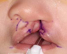
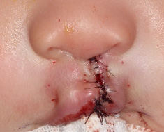
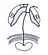
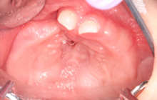
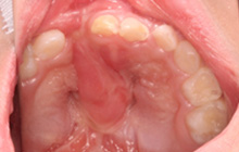
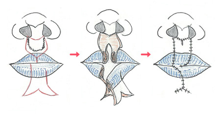
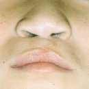
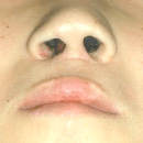
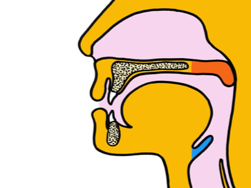
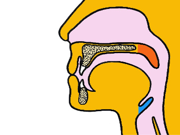
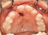
It was mentioned in detail earlier that cleft palate causes various problems. Palatoplasty is performed to solve these problems, but the purpose of this operation is not only to close the cleft but also to suture the splitted 
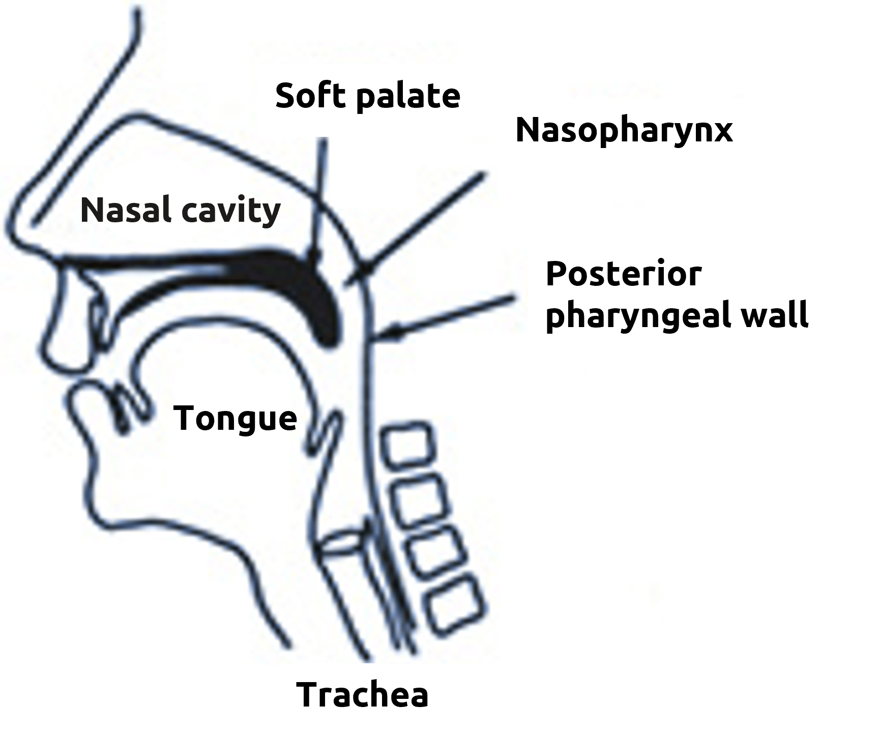
soft palate muscle (levator veli palatini muscle), return it to a normal position, and reduce the dilated rhinopharynx.

If cleft palate surgery is performed too early, it will impair the growth of the upper jaw. However, if the surgery is delayed too much, the habit of abnormal pronunciation (abnormal articulation) will develop. Therefore, in our department, surgery is performed at the age of around one year, when both of these conditions are satisfied fully, that is, before the baby starts active pronunciation exercise, and at the time when there is as little growth of the upper jaw as possible. However, in reality, the timing may change a little depending on the condition of the baby at that time.

Moreover, there are cases where the palate surgery is completed in one stage and cases where the surgery is performed in two stages. The reason for dividing the surgery into two stages is to improve the growth of the upper jaw as much as possible, but the optimal method changes depending on the condition of the mouth, so preoperative examination is performed to determine which method is better. Even in the case of the two-stage method, the first palatoplasty is performed around the age of 1 year, and the second surgical hard palate closure is performed around the age of 1 year and 6 months to 2 years. The surgery is performed under general anesthesia, and the length of the hospital stay is about 2 weeks.

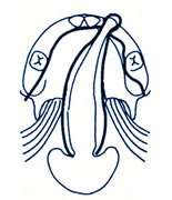
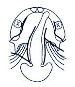
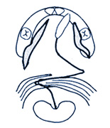
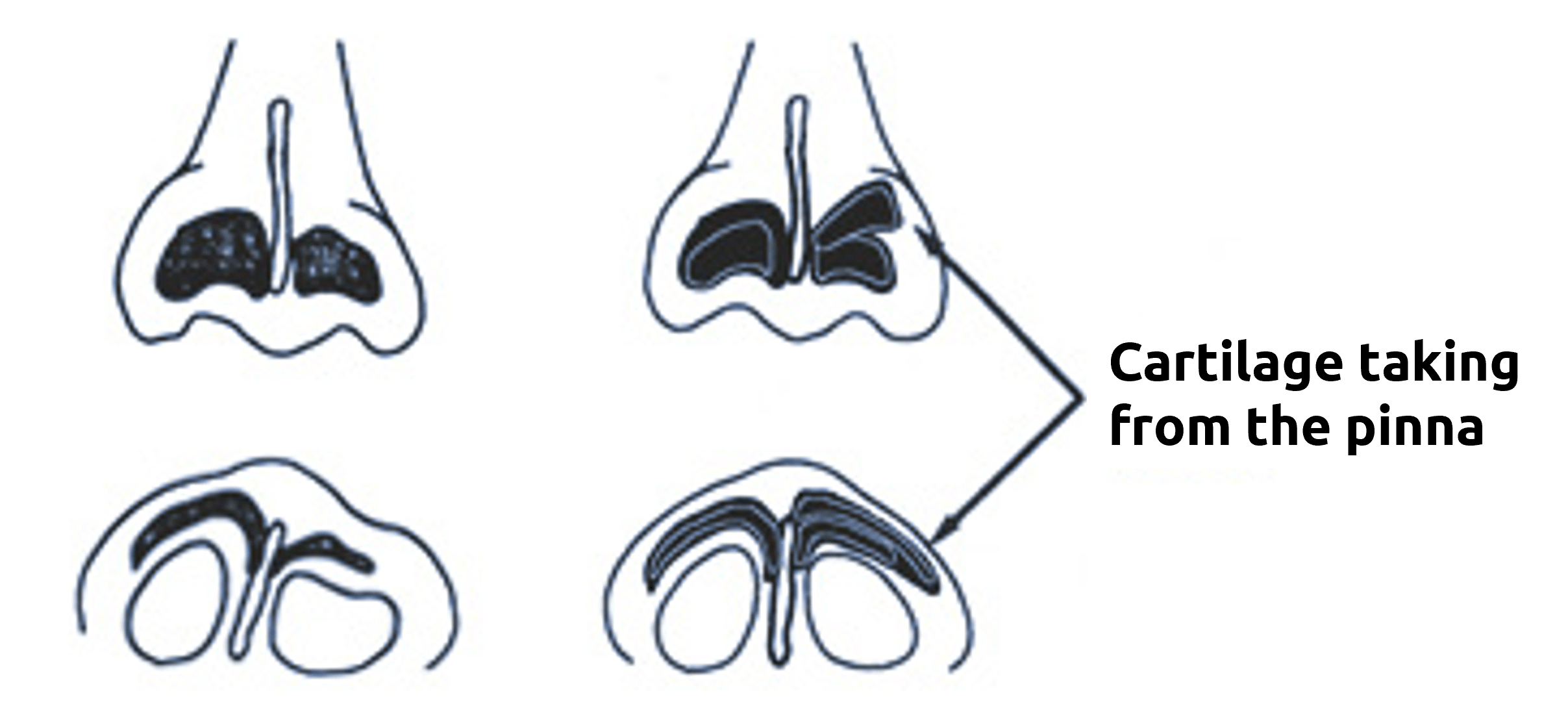
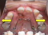
Regarding the surgical method, we select the optimal surgical method for each patient from the following two surgical methods based on the width of the cleft, the length of the soft palate, and the state of muscle development.

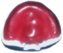
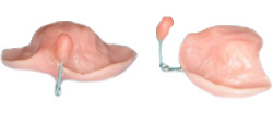
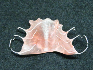
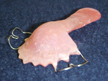
In both surgical procedures, the surgery causes partial bone exposure of the upper jaw, so the entire upper jaw is covered with a celluloid cover until a new mucous membrane is formed to protect the wound. The period of wearing it is about one week. Please eat soft meals for a while after discharge. After about 4 weeks after surgery, it is possible to return to the same diet as before surgery.

Additionally, since infants at this time put anything in their mouths, please be careful not to put any sharp objects (ball pens, chopsticks, etc.) into the mouth (please be especially careful until around 3 months after surgery).

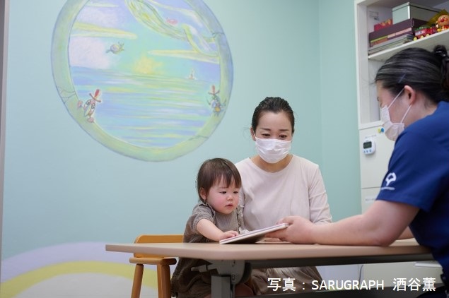
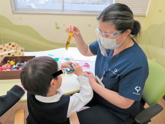
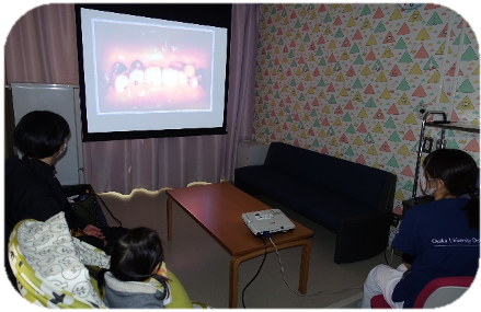
Regarding speech training, you will visit our division of Oral-Facial Disorders during the hospitalization to receive detailed explanations about future treatment (details will be described later).

1. Push back method

Push back method
Push back method

2. Furlow’s method

Furlow’s method
Furlow’s method
Before palatoplasty

Before palatoplasty

After closure of soft palate and posterior hard palate

After closure of soft palate and posterior hard palate

After closure of anterior hard palate

After closure of anterior hard palate

Preschool revision surgery

If the scars on the white lip area (the area between the red lip and the nose) are noticeable after cheiloplasty, or the shape of the red lips is irregular, or the nose is deformed, the patient will undergo corrective surgery before attending school. Since this is the time when the nose and upper jaw are growing, large-scale surgery may affect the growth of the face, so only relatively small surgery is performed.

Major surgery (e.g., nose formation due to transplantation of ear cartilage) is performed after the patient's growth and development are almost complete (14–18 years).

Bone grafting to the alveolar cleft and closure of the residual hole (hole in the upper jaw left after palatoplasty)

The bones of the alveolar ridge of the upper jaw (where the teeth are lined up, i.e., the gums) are lined up with not only teeth but also the base of the nose. Therefore, in patients with an alveolar cleft, the base and ala of the nose appear to be depressed. Additionally, because there is no bone that forms the base of the teeth, the eruption of the anterior teeth cannot be expected, and the teeth cannot be moved by orthodontic treatment. To solve such problems, our department perform surgery to transplant bone marrow collected from the ilium to this area around the age of eight.

In the case of patients with a cleft palate that extends to the alveolar region, the alveolar region is closed only by the mucosa during palatoplasty. However, if the width of the fissure is very large, it may be difficult to close the alveolar region completely, and after surgery, a small hole (residual hole) that connects to the nasal cavity may remain in the alveolar region. Even if there is no hole after surgery, a hole may be formed around the alveolar area by performing orthodontic treatment, lateral enlargement of the upper jaw. If this hole is large, the water in the mouth will leak to the nose, causing various problems such as difficulty using a straw. When producing sounds, the pronunciation may be obscured because there is not enough air in the mouth. Therefore, we are trying to solve such problems by performing surgery to close this hole (residual hole closing surgery) together with bone grafting to the alveolar region mentioned previously.

Surgery for bone grafting to the alveolar region is performed at an appropriate time from the age of 5 in consultation with the orthodontic department. Bone is collected from the ilium or mandible according to the required amount. This cancellous bone is a sherbet-like bone that exists inside the bone, and a part of the outer hard bone (cortical bone) is removed for collection, but the hard bone is replaced after collection, so there is almost no deformation of the ilium. Even if the collection from the ilium is performed, it is possible to walk within 3 days from the day after the operation. The collection from the chin cannot be performed unless the mandibular anterior teeth have been replaced with permanent teeth.

If the hole in the palate is large, the mucous membrane of the tongue may be transplanted to close the hole (tongue flap transplantation), and these operations completely eliminate the alveolar fissure (jaw cleft).

Closure of the residual hole

Correction of the external nose (external nose correction)

In patients with a unilateral cleft, the growth of the cartilage that forms the nose differs between the right and left sides, and the difference may be noticeable as the body grows. In patients with a bilateral cleft, the central part of the nose, especially the tip of the nose, may be stunted and the nose may be low. We perform such external nasal morphological surgery from the age of 14 when the growth of external nasal cartilage is almost complete (this period varies between men and women).

The surgery focuses on restoring displaced nasal cartilage with the aim of making the shape of the nose symmetrical, but if significant cartilage growth impairment is noted, we will transplant the auricular cartilage and the mandibular chin bone. The cartilage of the auricle is collected from the back of the ear, so the scar is unnoticeable. Additionally, since the cartilage is collected in a window shape, the shape of the ear does not change after surgery.
Additionally, although some facilities perform silicon transplantation for the formation of the nose, we do not use silicon as the adverse effects of transplanting artificial materials cannot be ruled out.

After surgery, it is necessary to wear a silicone nasal plug (Retina) for 3 to 4 months to prevent relapse of the morphology.

Figure: Cartilage collected from the auricle
orrection of the external nose (external nose correction)

Lip correction surgery

When a child is 14–16 years old and has passed the growth period, the lips and nose may change from a child’s shape to an adult’s shape, resulting in a marked distortion. If there is concern about this kind of distortion, the final surgery to shape the lips is performed at this time, similar to the above-mentioned revision surgery for the external nose.

The surgery method is completely different for each patient and may include repairing only the red lip, repairing scars on the white lip, or using external rhinoplasty to correct the entire lips. Small corrections may be performed outpatiently, but if relatively large surgery is required, surgery is performed under general anesthesia after hospitalization. After the surgery, it may be required to apply tape to make the scars less noticeable, just like after the lip plastic surgery when the patient was a baby.

There is also a surgical procedure to transplant the tissue of the lower lips to the upper lips (inversion flap transplantation [Abbe’s flap transplantation]). This surgery is performed for patients with bilateral cleft lip whose upper lip is extremely poor growth and whose lower lip is relatively prominent or for patients with severe scarring.

Secondary cleft lip repair

Secondary cleft lip repair

Before operation

Secondary cleft lip repair

After operation

Abbe’s flap in secondary cleft lip repair

Abbe’s flap in secondary cleft lip repair

Before operation

Abbe’s flap in secondary cleft lip repair

After operation

Abbe’s flap in secondary cleft lip repair

Before operation

Abbe’s flap in secondary cleft lip repair

After operation

Speech treatment for cleft lip and palate

Speech issues

Cleft palate (including after surgery) may cause specific language problems, and these problems are generally when pronouncing words:

  • 1
    Problems with how to use (operate) the mouth
  • 2
    Problems of nasal air emission and excessively nasal voice
  • 3
    Other problems that are unrelated to cleft lip and palate
    (developmental issues, congenital hearing disorder, etc.)

There are various symptoms for each of problems above. Especially in the case of problems of (1), symptoms such as "specific sounds being replaced by different sounds" and "baby-talk" can be observed. Furthermore, in the case of problems of (2), symptoms such as "talking though nose" and "mumbled, unclear speech" are often observed. However, speech may become unclear by any of the problems above.

Symptoms and their causes may be related to just one of these problems or may be related to multiple problems and may vary from person to person. Therefore, it is important to confirm the cause of speech problems and take appropriate measures in address to each cause.

Relationship with Department of Oral-Facial Disorders

1 First relationship

This department is responsible for speech management and treatment. Dentists and speech therapist from this department will see the patients and families after the first maxillary surgery (palatoplasty).Since this surgery is performed around the age of one at our hospital, majority of the children have not yet started to talk at this point.

By being involved from this time and watching over the development of speech, we can detect speech problems at an early stage and carefully judge whether there is a need for speech therapy and/or treatment or not. Dentists and speech therapist will explain the symptoms and treatment recommendatior in detail to the patients and families.

Relationship with Department

2 Speech training

If the speech problem falls under (1) or (2) above, speech therapy may be recommended. Speech therapists will teach the patients (and families as well) how to pronounce the speech sounds correctly. Home training is usually a great way to enhance patients’ speech.

Speech therapists make the recommendatior of the optimal program, including the start and frequency of therapy sessions for each case. For most of the cases, therapy starts around the age of 4 to 5, before attending school, and the frequency of therapy sessions are about once or twice a month.

Speech training

3 Treatment with the oral appliance

If the speech problem falls under (2) above, either there may be a hole remaining in the upper jaw (residual fistula) or there may be a problem with the function or length of the velum (soft palate) (nasopharyngeal insufficiency (Fig. 1)).

We will create a removable oral appliances to temporarily close the fistula (palatal plate) or to support the velopharyngeal insufficiency (speech aid: Fig. 2) for velopharyngeal insufficiency. By using a device made by dentists, speech therapists will provide speech therapy.

Figure1: Velopharyngeal insufficiency
鼻咽腔閉鎖機能不全

When pronouncing many sounds, the soft palate (orange) lifts and closes between the nose and throat.

鼻咽腔閉鎖機能不全

However, if there is velopharyngeal dysfunction, the soft palate does not move well and voice and air leak into the nose, resulting in an unclear pronunciation.

Figure2: Examples of oral appliances and speech aid
Examples of oral appliances and speech aid

Oral appliances:The device closes the residual fistula and prevents voice and air from leaking into the nose.

Examples of oral appliances and speech aid

Speech aid: An example of the device is called a palatal lift prosthesis (PLP). It helps lift the soft palate and improves velopharyngeal closure.

4 Surgical treatment

In the case of persons who need speech aid for achieve good speech, secondary speech surgery can be used to improve their speech instead of using the device. This procedure is called pharyngeal flap surgery. There are several methods for this surgery, and the age at which it is performed may vary from facility to facility.
At our hospital, we choose to wait until the upper jaw completes its growth, usually around high school age, before this surgery. However, when osteotomy (jaw surgery to improve the dental bite) is planned, pharyngeal flap surgery will take place after the jaw surgery.

5 Others

If the speech problem involves the problems discussed in (3) above, we will help and support the patients and families in coordination with other facilities that are specialized in the relevant issues.

Orthodontic treatment of cleft lip and palate

Why is orthodontic treatment necessary?

Children with cleft lip and/or palate (CLP), may have complex problems such as misalignment of teeth and jaw, deformities of the upper jaw (maxilla), and missing /poorly shaped teeth.

In most children with CL/P, orthodontic treatment is required for ensuing harmonious and appropriate dental and facial form. Orthodontic treatment is also recommended to be performed at the appropriate time in accordance with the individual conditions.

Why is orthodontic treatment necessary?

Figure: A midfacial deficiency, related to undergrowth of the maxilla, is caused by the effect of cleft and its surgery.

Why is orthodontic treatment necessary?

The flow of orthodontic treatment

1 First visit and examination (around 4-5 years old)

An initial consultation is performed at first visit. After the initial consultation, an appointment of examinations will be taken. Orthodontic examinations include intraoral photography, facial photography, dental models, intraoral scan, X-ray, CT, and 3dMD. Based on the examinations, a customized treatment plan for the patient is developed by the multidisciplinary team involving specialists from orthodontics, surgery, speech therapy, pediatric dentistry and prosthetic dentistry.

Orthodontists are involved with guidance of growth and development regarding the face and dentitions, in order to establish the harmonious and appropriate oral function and facial appearances among the children with CL/P.

Figure: Face/head photography using 3dMD and 3D data analysis

Face/head photography using 3dMD and 3D data analysis

Figure: Superimposition of CT and 3D facial image

Superimposition of CT and 3D facial image

Figure: Intraoral scan

Intraoral scan

2 Diagnosis

Orthodontists explain the diagnosis, recommended orthodontic treatment options, and implications. Orthodontists, patient, and their parents discuss and decide the final treatment plan.

Diagnosis

3 Orthodontic treatment for children

Expansion of the upper jaw (maxilla)

The maxilla is frequently collapsed in transverse dimensions in the children with CL/P. The maxilla may be expanded in order to correct the collapsed maxillary arch. The maxillary expansion sometimes grows the oronasal fistula, which may cause the problem such as liquids escaping through the nose. An obturator is sometimes used to close off the fistula with the orthodontic appliances.

Before orthodontic treatment

Before orthodontic treatment

Expansion of upper dental arch

Expansion of upper dental arch

Quad Helix Appliance

Protraction of the maxilla

An undergrowth of the maxilla often occurs in the children with CL/P. A reverse headgear (protractor) may be used to protract the maxilla and promote the growth of maxilla.

Lingual arch may be also used to improve an anterior crossbite, caused by mispositioned the upper incisors.

Maxillary protraction headgear

Maxillary protraction headgear

Lingual arch

Lingual arch

Bone grafting of the alveolar cleft

Bone grafting of the alveolar cleft is usually done during the period of 6-10 years before the canine erupts.

The optimal time for bone grafting depends on individuals’ conditions, such as the size/type of the cleft, teeth, and the progress of orthodontic treatment. After bone grafting, the alignment of incisors may be done.

顎裂部骨移植術

4 Orthodontic treatment after the adolescent growth spurtDefinitive orthodontic treatment begins after the permanent teeth have erupted or the growth of the jaw has completed.

Orthodontic treatment using fixed appliances

As the growth of lower jaw continues until around ages 16-18 years old, the occlusion and jaw relations may also change with the growth. After the permanent teeth have erupted or the growth of the jaw has completed, definitive orthodontic treatment using fixed appliance begins.

In some case, the surgical intervention may be required to optimize the occlusion and to improve the facial appearance.

装置を用いた治療

Protocol of orthognathic surgery
  1. Preoperative orthodontic treatment

    Preoperative orthodontic treatment is performed to align the teeth so that the occlusion fit correctly after surgery is performed.

  2. Jaw surgery (orthognathic surgery)

    Jaw surgery will reposition or realign the jaw to correct the discrepancies between the jaws. The jaw bone will be moved into the planned positioned and fixed with plates and screw. A splint and tight orthodontic elastics may be kept after surgery. They help the stabilization of the repositioned jaws.

  3. Postoperative orthodontic treatment

    After jaw surgery, postoperative orthodontic treatment is performed to finalize dental occlusion in the new skeletal relationships.

Orthognathic-surgical treatment

Orthognathic-surgical treatment

Before treatment Just before surgery After surgery

Profile of post treatment

Profile of post treatment

Before surgery After surgery

Several types of jaw surgeries for correcting underbite
  • Maxillary osteotomy:

    Maxillary osteotomy can correct a midfacial retrusion.

  • Mandibular osteotomy:

    Mandibular osteotomy can correct a protruded chin.

  • Two jaw surgery:

    Two jaw surgery can correct both of a midfacial retrusion and a protruded chin.

  • Maxillary distraction osteogenesis:

    Maxillary distraction osteogenesis is a surgical procedure that expands or extends bone in the upper jaw. In Maxillary distraction osteogenesis, the slow movement of two apart bony segments can create new bone and fill in the gap.

Mandibular osteotomy

Mandibular osteotomy SSRO

SSRO

Mandibular osteotomy IVRO

IVRO

2 Jaw orthognathic surgery

2 jaw orthognathic surgery + SSRO

Maxillary osteotomy + SSRO

Segmental maxillary osteotomy + SSRO + genioplasty

Segmental maxillary osteotomy + SSRO
+ genioplasty

  • Maxillary anterior segmental distraction osteogenesis (MASDO):

    MASDO is a surgical procedure for patients with a severely midfacial retrusion due to the undergrowth of upper jaw. MASDO is effective for improving the facial profile without velopharyngeal problems and regaining the alveolar space.

上顎骨前方部仮骨延長術(MASDO)

The distracter is set by surgery.

MASDO is a new technique developed in first department of oral maxillofacial surgery.

Retention:

Retention is the final stage of orthodontic treatment. The purpose of retention is to maintain the teeth in their corrected positions after the active treatment.

保定

Oral hygiene management in children with cleft lip and palate

In pediatric dentistry, thorough oral hygiene during childhood, especially prevention of dental caries, is kept in mind. Additionally, we are focusing on treating caries that have developed as soon as possible while inhibiting their progression.

Oral hygiene management

A lecture “The Story of Oral Health” using slides

Oral hygiene management

Dietary guidance for parents

Based on the results of surveys to date, it is well known that children who visit the hospital for the treatment of cleft lip and palate are more likely to have dental caries. In particular, the upper front teeth that erupt near the cleft tend to develop caries, and it is necessary to think carefully about the prevention of caries from the time when the front teeth start to erupt.

Therefore, in pediatric dentistry, we focus on tooth brushing instructions by the dentist in charge and dental hygienist. The points regarding how to brush the upper front teeth, which are especially prone to tooth decay, are shown in the following:

A case of the upper front tooth caries next to the alveolar cleft

A case of the upper front tooth caries next to the alveolar cleft

  • 1
    Be careful not to let the tip of the toothbrush come into contact with soft tissue such as the gums.
  • 2
    When brushing the labial side of the upper front teeth, the index finger is inserted slightly from the side and the entire pad of the finger is used to lift the upper lip.
  • 3
    For the backside of the upper front teeth, which is particularly difficult to brush, a one-tuft brush that has a small brush part and allows pinpoint brushing is recommended to be used.
One tuft brush

One tuft brush

Prevalence of dental caries in 4-year-old children who go to the hospital for the treatment of cleft lip and palate

It has been found that, in children who go to the hospital for treatment of cleft lip and palate, the upper front teeth that grow near the cleft are likely to develop caries, but the lower back teeth are also likely to develop caries. Therefore, it is necessary to pay close attention to the entire teeth in the mouth.

It is generally known that children who have many caries when they have deciduous teeth will have many caries even if they are replaced with permanent teeth. This is partly because the number of dental caries bacteria in the mouth is determined in early childhood.

Additionally, when the orthodontic treatment begins and an orthodontic appliance is placed in the mouth, toothbrushing becomes difficult, and the oral environment becomes even more prone to tooth decay. Therefore, it is very important to maintain a healthy oral condition by the time permanent teeth begin to erupt.

Deciduous teeth

Deciduous teeth are called in Alphabet, A, B, C, D, E.
Ex) Upper right A, Lower left D.

Prevalence of dental caries

Prevalence of dental caries

Although there are individual differences depending on the condition of the child's mouth, regular examinations are conducted every 3 to 4 months.

In regular check-ups, the following are performed.

  • Check for dental caries

    By performing regular medical examinations, it is possible to detect cavities and respond to them at an early stage.

  • Guidance on tooth brushing

    We provide tooth brushing guidance according to the age and oral condition of each child. We will focus on how to select the right toothbrush and how to brush the teeth that erupt near the cleft. We also give guidance to children so that they can brush their teeth by themselves when they get older.

  • Guidance regarding meals/between meals

    We provide dietary guidance and guidance on eating snacks according to the child's age and oral condition.

If desired, we provide preventive measures for dental caries such as fluoride application and fissure sealant.