We are working on "development and regeneration of the skeletal system and orofacial structures."
![]()
Research
INDEX
- I. Understanding of cell fate specification processes in development and maintenance of the skeleton and teeth
- II. Study on the role of CTRP3/C1qTNF3
- III. Elucidating the molecular mechanisms of axial and appendicular skeletal morphogenesis
- IV. Neurobiological studies on oral sensations
I.Understanding of cell fate specification processes in development and maintenance of the skeleton and teeth
We pursue two scientific interests with a particular focus on skeletal tissues (bone and cartilage) and teeth: (1) basic understanding of the molecular mechanisms underlying development and maintenance of tissues and organs, and (2) the development of fundamental technologies for tissue regeneration and repair. We aim to understand cell fate determination processes in terms of gene regulation and epigenome dynamics and to provide proof-of-concept of the genome-inspired strategy for tissue regeneration and repair. We take advantage of a combinatorial approach including morphology, developmental biology, molecular biology, stem cell biology, mouse genetics, bioengineering, genome-wide analyses, and bioinformatics.
A)Epigenome dynamics and gene regulatory networks during cell fate specification processes
We seek to understand the mechanisms of cell fate specification, cell differentiation, and cell maturation during skeletal development, with a particular focus on epigenome dynamics and genomic targets of transcription factors. We take advantage of genome-scale data on gene expression, the epigenome, and transcription factor binding sites in order to investigate the regulatory landscape of the genome, which determines the phenotype of osteoblasts and chondrocytes. A series of studies has revealed transcriptional networks that mediate pluripotency and the differentiation of pluripotent stem cells (Stem Cells 31:2667, 2013), epigenome dynamics and the mode of action of master transcription factors in osteoblasts and chondrocytes, and their mediating gene regulatory network (Cell Reports 12:229, 2015; Developmental Cell 37:238, 2016; Development 143:3012, 2016; Trends in Genetics 32:774, 2016; Cell Reports 40:111315, 2022; Cell Reports 42:112276, 2023). With respect to signaling pathways regulating skeletal formation, we have focused on Hedgehog (Hh) signaling. We have identified roles of Hh signaling in specification of bone-forming osteoblasts and homeostasis of bone tissues (Developmental Cell 14:689, 2008; Journal of Biological Chemistry 287:17860, 2012; Journal of Biological Chemistry 288:9924, 2013; PLoS ONE 9:e109597, 2014). A fuller understanding of the cell fate specification processes during skeletal formation, we believe, will enable the development of novel therapeutic strategies.
B)Tissue-development modeling systems using pluripotent stem cells
Pluripotent stem cells (PSCs)—i.e., embryonic stem (ES) and induced pluripotent stem (iPS) cells—have the ability to self-renew and differentiate into all of the lineages present in the body, and are thus a promising tool for developmental biology and regenerative biology. The reproduction of organogenesis and metabolism in vitro may contribute not only to drug discoveries and regenerative medicine, but also to understanding of disease pathophysiology and of the molecular mechanisms underlying tissue formation and maintenance. We have therefore been developing protocols for directing mouse and human PSCs toward osteoblasts under defined conditions using only small molecules, while recapitulating in vivo osteoblast development (Stem Cell Reports 2:751, 2014; Regenerative Therapy 14:19, 2020; Cell Reports 42:112276, 2023; Regenerative Therapy 24:536, 2023). We are also attempting to apply these protocols to the generation of PSC-derived bone-like tissues, in which distinct cell populations regulating bone formation and maintenance (osteoblasts, osteocytes, and osteoclasts) function on culture dishes in a three-dimensional manner (Science Advances 3:e1602875, 2017).
C)Bioactive factors that induce bone/cartilage formation
Skeletal formation is regulated by various signaling pathways and transcription factors. The manipulation of key pathways and/or factors would enable us to not only induce skeletal formation and regeneration, but also suppress the progression of skeletal degeneration. Thus, we are carrying out molecular-biological and mouse genetic studies to elucidate the role of osteogenic and chondrogenic signaling pathways as well as the mode of their actions. Based on these basic findings, we are also working on the identification and application of bioactive molecules that induce osteogenesis and chondrogenesis. In this context, we have been focusing on: (1) hedgehog (Hh) signaling-mediated cell-fate specification during osteoblast development (Development 131:1309, 2004; Journal of Biological Chemistry 282:17860, 2012; Journal of Biological Chemistry 288:9924, 2013), and (2) transcriptional regulation mediating osteoblast differentiation and maturation (Developmental Cell 14:689, 2008; PLOS ONE 9:e109597, 2014). These studies have further extended to the development of a small molecule-based treatment for bone fractures using the Hh signaling-activator molecule SAG (Biochemical and Biophysical Research Communications 479:772, 2016) and a bone-regenerative therapy using SAG-loaded calcium phosphate artificial bones (Biomaterials 34:5530, 2013). We also identified the novel osteogenic compound, helioxanthin analogue TH, which is commercially available (Biochem Biophys Res Commun 357:854, 2007; https://www.tcichemicals.com/eshop/ja/jp/commodity/M3085/)
| Collaborators |
|---|
| The University of Tokyo |
| Nagasaki University |
| Tohoku University |
| Tokyo Dental College |
| Tokyo Medical University |
| Showa University |
| University of Connecticut Health Center |
| Pharma Foods International Co., Ltd. |
| Institute for Theoretical Medicine, Inc. |
II. Study on the role of CTRP3/C1qTNF3
CTRP3/C1qTNF3, initially identified as a growth plate cartilage-derived 26-kDa secretory protein, is a member of a family of adiponectin paralogs, known as C1q and tumor necrosis factor (TNF)-related proteins (CTRPs). Adiponectin is one of the adipocytokines that has been extensively studied. Today, the CTRP family consists of about 15 members. Although the functional receptors for CTRPs have not yet been recognized, CTRP3/C1qTNF3 has multiple effects on lipid and glucose metabolism, inflammation and musculoskeletal disorders. We generated Ctrp3 knockout (KO) mice and have been studying their phenotypes to reveal novel physiological and pathophysiological functions of CTRP3/C1qTNF3.
III. Elucidating the molecular mechanisms of axial and appendicular skeletal morphogenesis
Skeleton is indispensable organ for animals living under gravity. We are trying to understand the molecular mechanisms of skeletal morphogenesis based on mouse genetics together with cell and organ culture-based approaches.
- 1.
- Craniofacial structure is quite complex, and its normal development needs to be precisely regulated by the timely crosstalk between number of cell populations. At the same time, this mechanism should be relatively simple to be “robust”. One of the ways to make it simple is to educate small number of the leader cell populations and make rest of the cells to just “follow the leader”. We are trying to identify the profiles of these strong leader cells (Michikami et al., 2012; Nomir et al., 2016; Abe et al., 2021).
- 2.
- We are able to move as bones have joints between adjacent ones. Huge progress has been made for the molecular understandings of digit and limb joints. In contrast, the mechanisms of shoulder, hip, and temporomandibular joints are less understood. It is known that the cell origins of shoulder and hip joint differs (shoulder joint have considerable amount of neural crest cell population in addition to sclerotome and lateral plate mesoderm derived cells). Also, the crucial molecules involved in these two joint developments seem to differ. Currently, we are analyzing in detail a novel gene hypomorph mice which display severe hip joint development anomaly.
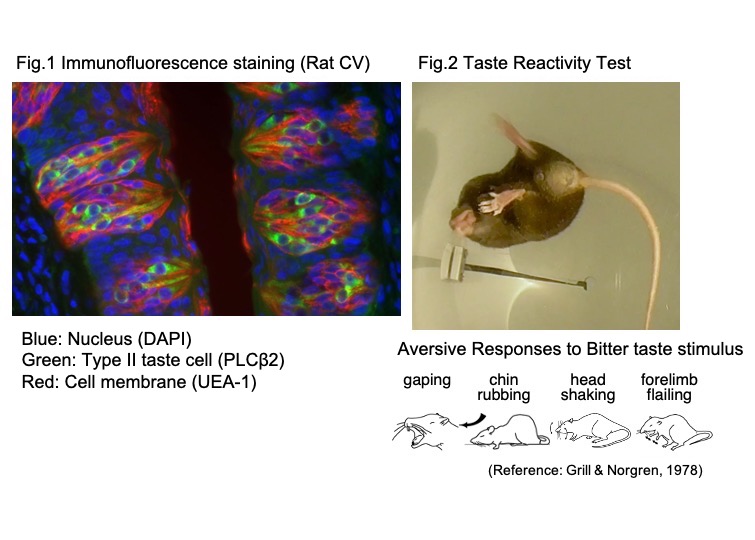
IV. Neurobiological studies on oral sensations
Our mission in dentistry is to elucidate how oral functions should be recovered and maintained. The mastication of foods is accompanied by the process of sensory organs such as taste, somatosensory, and temperature receptors. These sensory produce the feeling of “deliciousness” and “pleasantness.”
Tastes of foods consist of five basic tastes: sweet, salty, umami, sour, and bitter. Each basic taste has a biological meaning: sweet, salty, or umami is a signal of calorie, mineral, or amino acids, respectively, which are essential for humans and animals. On the other hand, sour and bitter tastes indicate harmful substances in spoiled and contaminated foods. The taste substances bind the receptors on taste cells in taste buds expressed on the tongue, soft palate, pharynx, and larynx. Excitation of the taste cells is transmitted to the brain via taste nerves.
In dental hospitals, the number of patients with taste disorders is increasing, which is thought to be related to the increasing population of aged people. The taste disorders declining sensitivity are possible to shift preference for taste from weak to strong. It induces overconsumption of nutrients which is a cause of lifestyle disease.
To clarify the mechanisms underlying the aging-related decline of taste function, we are conducting research at the level from genes to a whole animal. Our present studies are as follows: 1) decrease in body zinc level is suggested to be a critical cause of taste disorders. Thus, we use animal models showing taste disorders under low zinc conditions or promotion of aging to investigate the effect of aging on the anatomy and functions of taste-related peripheral and central nervous systems and animals’ behaviors. 2) Recent studies have suggested that taste receptors are expressed in the oral cavity and other peripheral organs. We are now focusing on the age-related changes in the function of the extra-oral taste cells.
We expect these studies to produce findings contributing to the development of preventive and treatments for diseases and establish a world where everyone can enjoy eating and drinking healthily.
Banner


Dept of Tissue and Developmental Biology
Graduate School of Dentistry
Osaka University
1-8 Yamada-Oka, Suita, OSAKA 565-0871, JAPAN
TEL +81-6-6879-2874
FAX +81-6-6879-2875